
Figure 3.
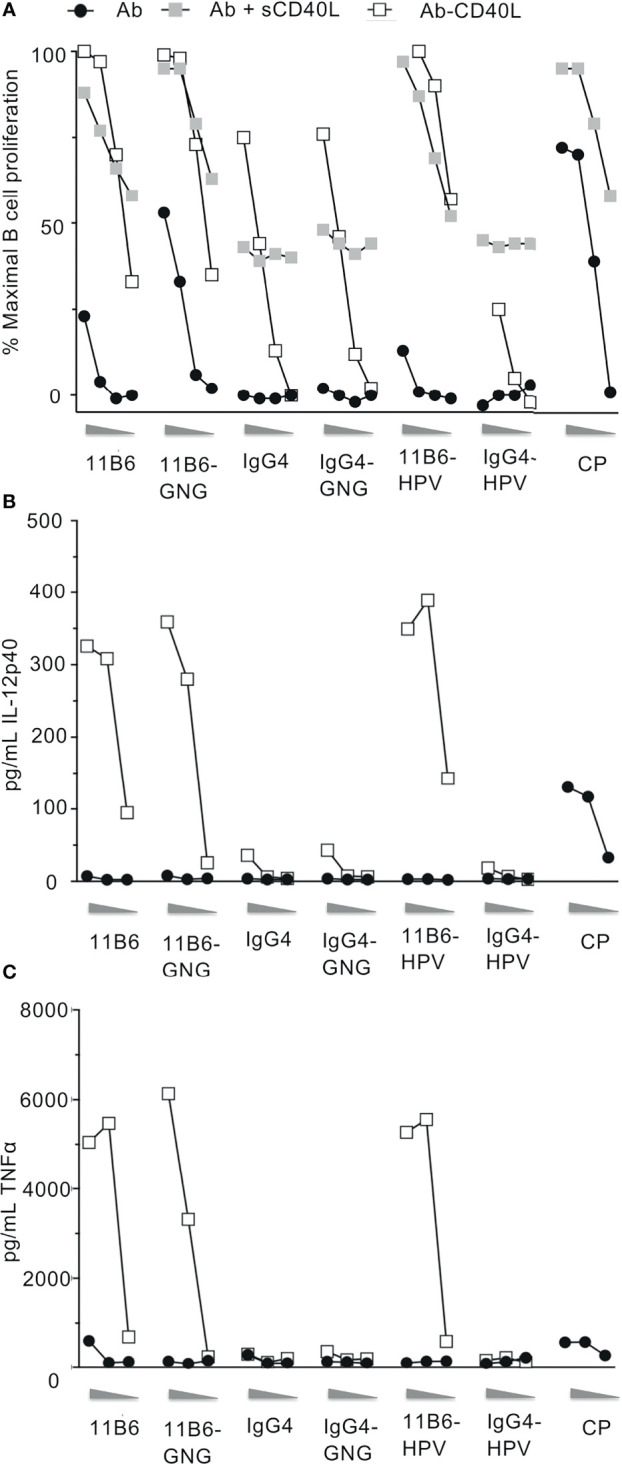
Anti-CD40 11B6-CD40L fused to HIV-1 Gag p24, Nef, and Gag p17 or HPV16 E6/E7 antigens is highly active on human B cells and DCs. (A) Shows B cell proliferation in response to a dose range (shown left to right: 10, 1, 0.1, 0.01 nM) of the indicated antibody or antibody-antigen fusion protein; curves with gray filled square symbols are responses doses to the indicated antibody or anti-CD40 11B6-antigen fusion protein in the presence of 1 μg/ml (60 nM) soluble human CD40L or fused directly to CD40L. Data represent a single experiment normalized for maximum proliferation (95%) versus baseline proliferation without antibody (10%). (B, C) show MDDC cytokine secretion responses to a dose range (shown left to right: 10, 1, 0.1 nM) of each indicated mAb. Data show observed concentration values in pg/ml. The maximal values were IL-12p40, 359 ng/ml; TNFα, 6134 ng/ml. A dose range of the agonistic CP hIgG4 antibody is included for reference.