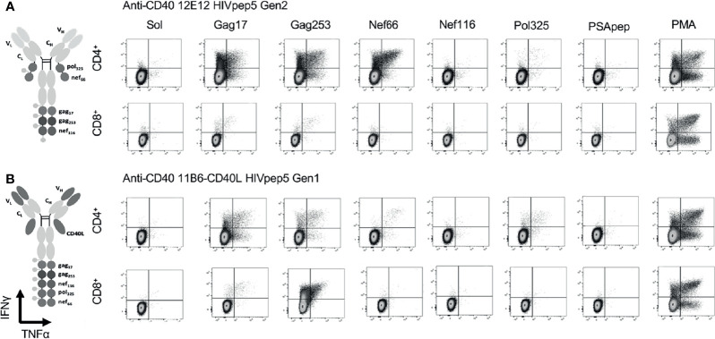
Figure 5.
CD40-targeted HIV5pep antigens with and without fused CD40L tested via in vitro expansion of HIV-1-specific T cells in HIV-1-infected donor PBMC cultures. HIV-1+ donor 1 PBMCs were cultured for 9 days with IL-2 and anti-CD40 HIV5pep fusion proteins (1 nM), followed by stimulation with long peptides specific for each of the five HIV-1 Gag, Nef, and Pol regions for 6 h with Golgi Stop + BFA, then analyzed by ICS. This is IHC data from one experiment with two of the four indicated proteins tested on one of the donors shown in Figure 6 . PSApep and sol indicate, respectively, non-relevant peptide and solvent only negative controls, and PMA represents polyclonal stimulation by Phorbol 12-Myristate 13-Acetate and Ionomycin. Minimal responses to specific peptides were observed in cultures expanded without vaccine stimulation (not shown) or with hIgG4-HIV5pep non-targeting control protein (7). (A) shows responses elicited by anti-CD40 12E12 HIV5pep Gen 2, and (B) shows responses elicited by anti-CD40 11B6-CD40L-HIV5pep Gen 1. Previous data show that Gen 1 versus Gen 2 HIV5pep configuration on anti-CD40 12E12 elicit identical ranges and extent of responses in similar experiments (14). Supplemental Figure 3 shows similar data for anti-CD40 12E12-HIV5pep Gen3 versus anti-CD40 11B6-CD40L-HIV5pep Gen3 responses in Donor 3 as a more direct comparison with matching Gen3 formats.

