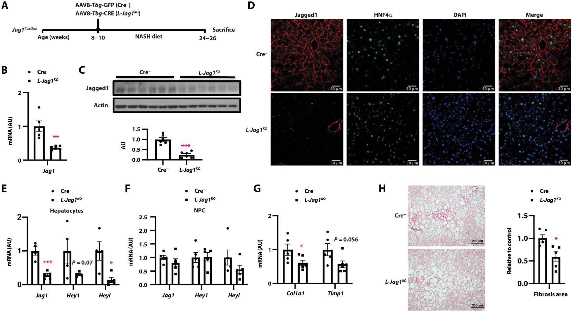
Fig. 3. Hepatocyte-specific Jag1 KO mice are protected from NASH-induced liver fibrosis.

(A) Adult Jag1flox/flox mice were transduced with either AAV8-Tbg-GFP (Cre−) or AAV8-Tbg-CRE (L-Jag1KO) and then fed NASH diet for 16 weeks before sacrifice. (B) Liver Jag1 mRNA and (C) protein expression, (D) representative image of Jagged1 (red) and HNF4α (green) staining, (E and F) Jag1 and Notch target expression in hepatocytes and NPC, and (G) gene expression markers of HSC activation. (H) Sirius red staining. n = 5 to 7 per group. *P < 0.05, **P < 0.01, and ***P < 0.001 as compared to Cre− mice by two-tailed t tests (two groups). All data are shown as means ± SEM.
