Figure 1.
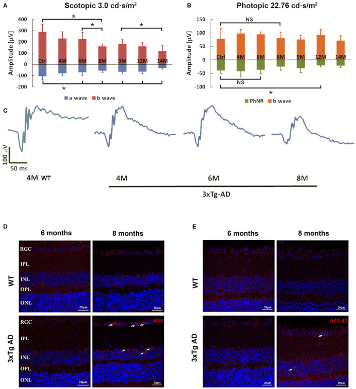
Flash-ERG and amyloid beta (Aβ) deposition in retinas of non-Tg control mice and 3xTg-AD mice at different ages. The scotopic retinal response (A) of 3xTg-AD mice started to decline at 6 months of age, followed by a rapid decrease from 6 to 8 months of age (A,C). *p < 0.05. Further significant decline in b wave changes can be detected between 6–8 months and 9–14 months. Significant reduction in a wave started at 6 months when compared with non-Tg controls. PhNR detected in the photopic response revealed a significant reduction at 12 months when compared with non-Tg controls (B). *p < 0.05; NS, no significant change. Typical ERG wave forms are shown in C. Intracellular Aβ deposition of 4G8 (D) and Aβ1-42 (E) were detected in the RGCL and INL of retinas of 3xTg-AD mice at 8 months only (arrows). No positive signal could be detected in the non-Tg controls or 6 month old 3xTg-AD retinas. Scale bar: 20 μm.

