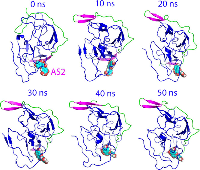
Figure 5.

Structure snapshots of MD simulations of the active form. Six individual structures of the first set of MD simulations for the active form of the dengue NS2B-NS3 protease complexed with myricetin at different simulation time points. For clarity, NS3 is displayed in the blue cartoon and NS2B with the β-strand colored in purple and the loop in green, as well as myricetin in spheres.
