Figure 10.

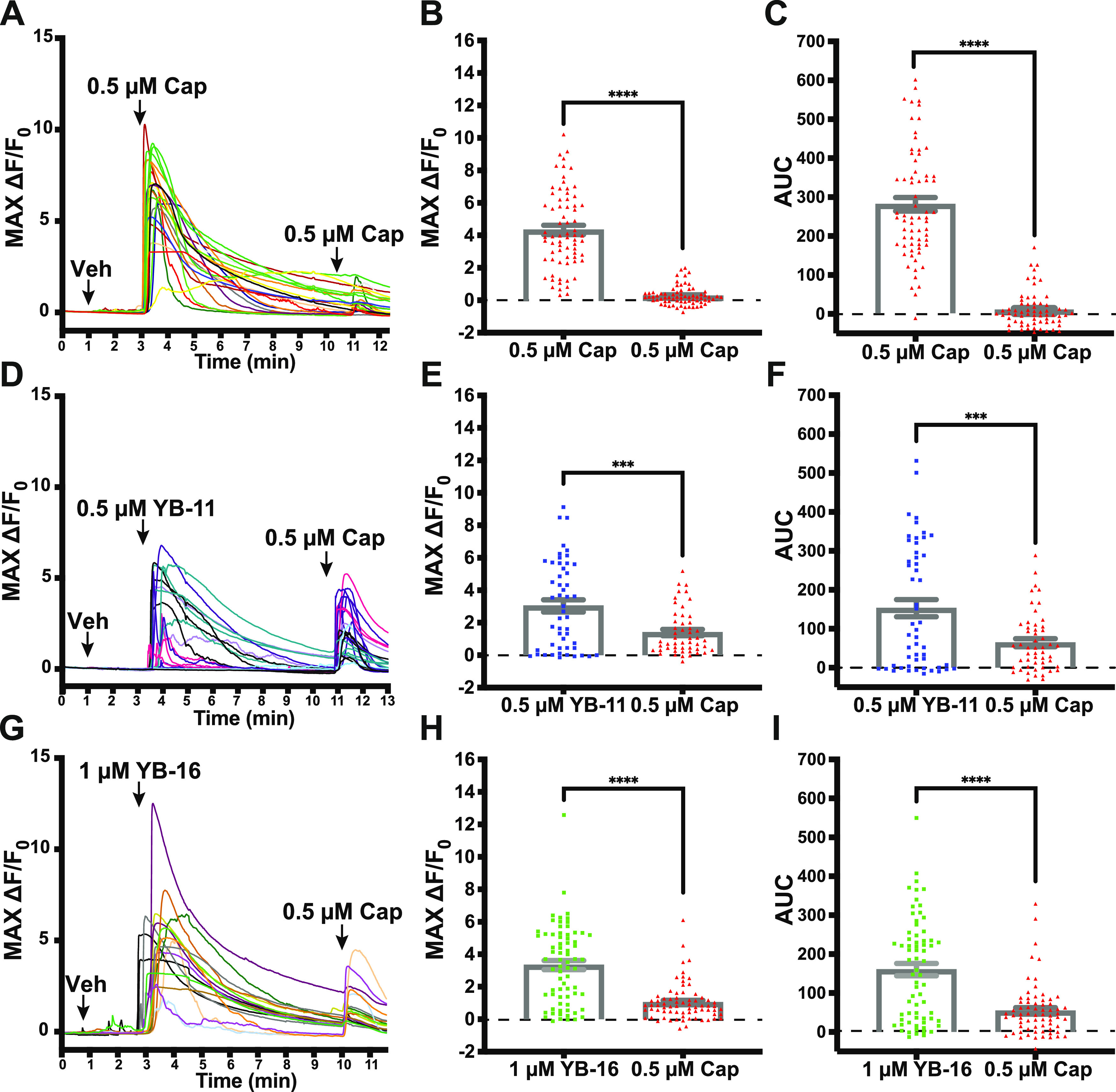
YB-11 and YB-16 on desensitized TRPV1-positive dorsal root ganglion as assessed with in vitro Ca2+ imaging. (A) Representative traces from an experiment, where dissociated DRG neurons were treated with capsaicin (0.5 μM) approximately 7.5 min before another treatment with capsaicin (0.5 μM). (B) The max Ca2+ signal from capsaicin was significantly reduced after the initial capsaicin response. (C) The area under the curve for the Ca2+ signal from capsaicin was significantly reduced after the initial capsaicin response. (D) Representative traces from an experiment, where dissociated DRG neurons were treated with YB-11 (0.5 μM) approximately 7.5 min before a treatment with capsaicin (0.5 μM). (E) The max Ca2+ signal from capsaicin was significantly reduced after the initial YB-11 response. (F) The area under the curve for the Ca2+ signal from capsaicin was significantly reduced after the initial YB-11 response. (G) Representative traces from an experiment, where dissociated DRG neurons were treated with YB-11 (1.0 μM) approximately 7.5 min before a treatment with capsaicin (0.5 μM). (H) The max Ca2+ signal from capsaicin was significantly reduced after the initial YB-16 response. (I) The area under the curve for the Ca2+ signal from capsaicin was significantly reduced after the initial YB-16 response. Paired t-test ***P < 0.001, ****P < 0.0001. Error bars in panels B, C, E, F, H, I are shown as mean ± SEM.
