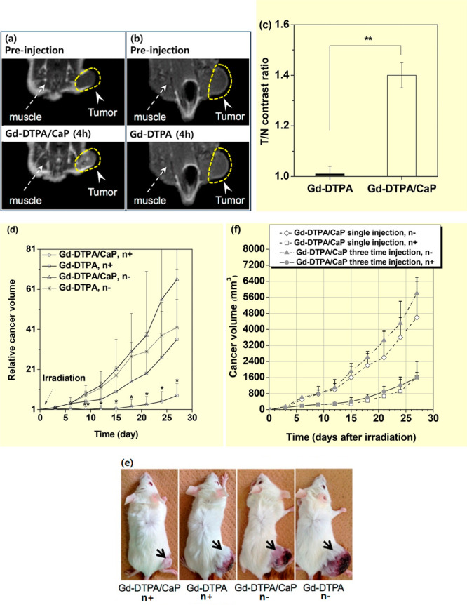
Figure 8.

T1-weighted MR images before and 4 h after intravenous injection of (a) Gd-DTPA/CaP nanocomposites and (b) Gd-DTPA. (c) Tumor-to-normal (T/N) tissue contrast ratios estimated from T1-weighted MR images in a and b (N = 3, p** < 0.01). (d) Relative cancer volume (Vday/Vday=0) of four C-26 cancer-bearing mouse groups as a function of days with and without thermal neutron beam irradiation (N = 4–5, p** < 0.01, *p < 0.05 from other groups). (e) Photographs of C-26 cancer-bearing mice taken on day 27 after thermal neutron beam irradiation. Adapted and reproduced from ref (14a). Copyright 2015 American Chemical Society. (f) Cancer volume of four C-26 cancer-bearing mouse groups as a function of days after thermal neutron beam irradiation. Reproduced with permission from ref (14b). Photograph courtesy of N. Dewi. Copyright 2016 Springer.
