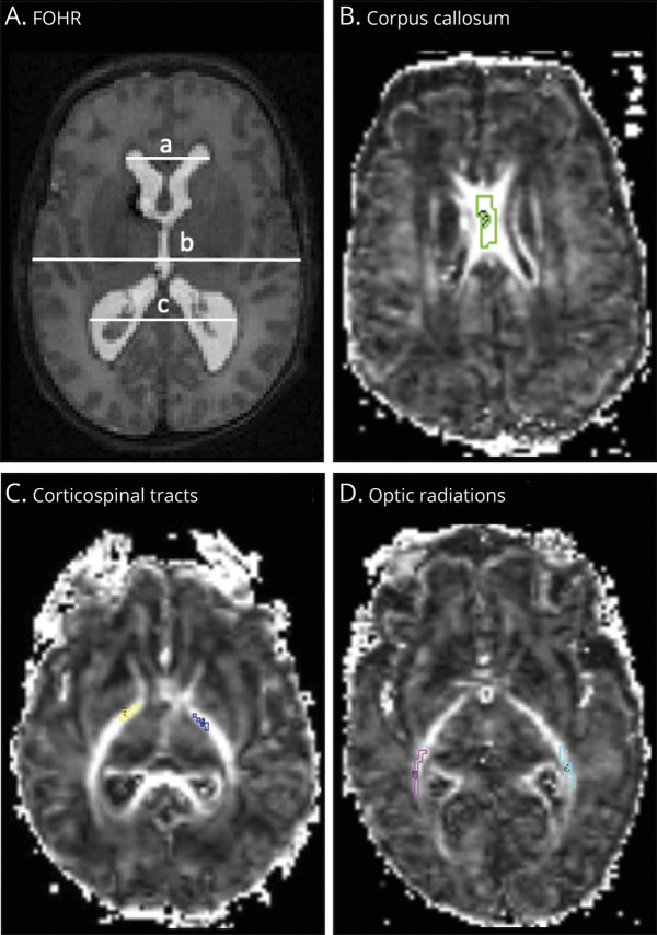
Figure 1. Representative Diffusion MRI.
Axial T2-weighted MRI scan (A) and contiguous axial fractional anisotropy maps (B–D) of infants born very preterm. Ventricular size was determined by the frontal-occipital horn ratio (FOHR), which measures ventricular size as the sum of the distance between the widest lateral walls of the frontal and occipital horns, divided by twice the widest biparietal diameter at the level of the foramen of Monro. (A) FOHR = (a+c)/2b. On the fractional anisotropy maps (B-D), segments of the white matter bundles of interest within the corpus callosum, bilateral corticospinal tracts, and bilateral optic radiations are demarcated.

