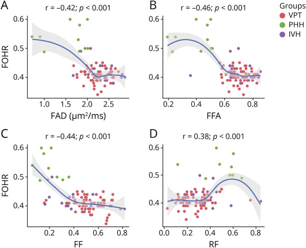
Figure 4. Ventricular Size–Diffusion MRI Correlations.
(A–D) Increase in ventricular size correlates with poorer diffusion MRI measures in the corpus callosum. As ventricular size increases, a decrease in white matter fiber axial diffusivity (FAD), fiber fractional anisotropy (FFA), and fiber density (fiber fraction, FF) and an increase in cellular infiltration (restricted fraction, RF) were observed. Ventricular size was obtained using the frontal occipital horn ratio approach (FOHR). IVH = intraventricular hemorrhage; PHH = post-hemorrhagic hydrocephalus; VPT = very preterm.

