Figure 4.

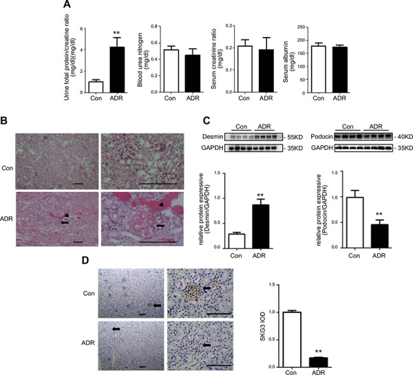
SGK3 protein expression was inhibited in mice with ADR nephritis. An intravenous injection of ADR (10.5 mg/kg) was used to induce nephritis. A) Plasma BUN, creatinine, albumin, and the urinary albumin/creatinine ratio were measured in mice after treatment with ADR. B) PAS staining demonstrates differences between control and ADR treated mice. Representative images from 7 mice in each group are shown. Arrows indicate sclerotic glomeruli. Arrowheads indicate tubular casts and dilatation. C) Top: immunoblot analysis for desmin and podocin expression; GAPDH was used for normalization. Bottom: quantification of desmin and podocin by densitometry. D) Left: histochemistry staining of SGK3 was performed on 5‐μm paraffin‐embedded kidney sections. Representative images from 7 mice in each group are shown. Arrows indicate glomeruli. Brown staining represents expression of SGK3. Right: computerized morphometric quantification of SGK3 staining in glomeruli in control and ADR mice. Data are given as mean ± se (n = 7/group). **P < 0.01 vs. control group. Scale bars, 100 μm.
