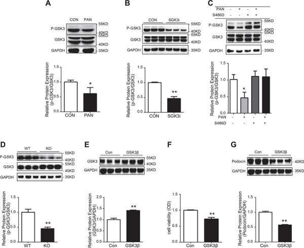
Figure 6.

P‐GSK3 regulated by SGK3 is related to the podocyte injury in vivo and in vitro. Top: immunoblot analysis for p‐GSK3 and GSK3 expression was performed. Bottom: quantification of p‐GSK3/GSK3 by densitometry. A) PAN treatment. *P < 0.05 vs. control group. B) SGK3 shRNA transfection. **P < 0.01 vs. control group. C) PAN treatment with S486D transfection *P < 0.05 vs PAN–/S486D– group. D) SGK3 KO mice. **P < 0.01 vs. SGK3 WT group. E) Top: immunoblot analysis for GSK3β expression; GAPDH was used for normalization. Bottom: quantification of GSK3β by densitometry. F) Podocyte viability was determined by MTT. G) Top: immunoblot analysis for podocin expression; GAPDH was used for normalization. Bottom: quantification of podocin by densitometry. OD, optical density. Data are means ± se of in 3 independent experiments. **P < 0.01 vs. control group.
