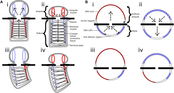
FIGURE 5.
Comparative diagram. (Ai–iv) shows the basic structure and motions of the tube foot of Asterias Rubens the common starfish, whilst (Bi–iv) shows the equivalent in our inflated actuator. Sections in red/solid are active muscles and DEAs, blue/striped are inactive, you can see the operation is inverted between the contracting biological muscle and our expanding artificial muscles. The operations of the ampulla and artificial ampulla are shown in figures (Ai–ii,Bi–ii) respectively, the arrows indicate the water/air flow.

