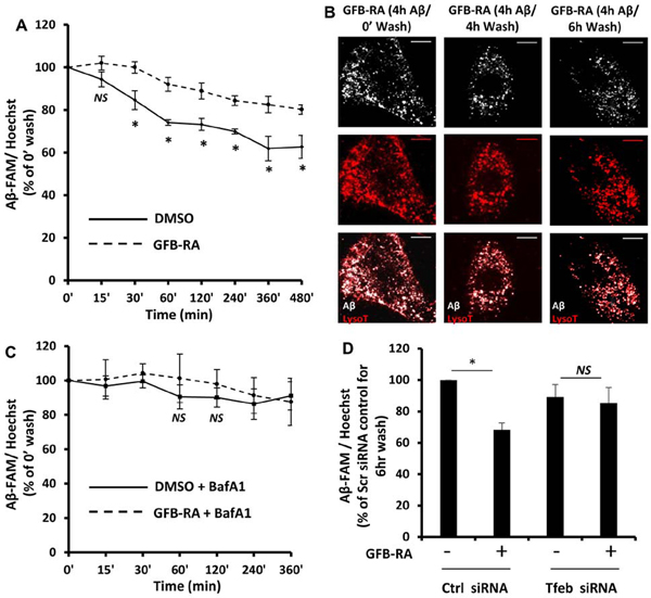
Figure 2. GFB-RA treatment enhances Aβ degradation in mouse primary astrocytes.
(A) Mouse primary astrocytes were treated for 24 hours with GFB-RA followed by incubation with 500nM FAM-tagged Aβ1–42 for 4 hours and allowed to grow in Aβ-free media for different time periods. Aβ degradation assay was performed as described in methods section. Data was represented a percentage change compared to unwashed control. (B) Mouse primary astrocytes treated with GFB-RA were incubated with 500 nM HF-Aβ1–42, washed for 4 or 6 hours, further incubated with 75 nM Lysotracker Red, and observed under microscope. Scale bar = 20μm. (C) Mouse primary astrocytes were treated with GFB and RA for 24 hours, followed by treatment with 100 nM Bafilomycin A1 for 45 min, followed by incubation with 500 nM FAM-Aβ, washed in Aβ free media for 6 hours and degradation assay was performed. Data is represented as percentage change with respect to unwashed controls. (D) Aβ degradation assay was done in mouse primary astrocytes which were either transfected with scrambled siRNA or Tfeb siRNA, prior to treatment with DMSO or GFB-RA. Data were compared to DMSO-treated, scrambled siRNA transfected controls. All data are mean ± SEM of three independent experiments run in duplicate. *P<0.05 and NS denotes not significant by one-way ANOVA followed by Dunnett’s multiple comparison test.

