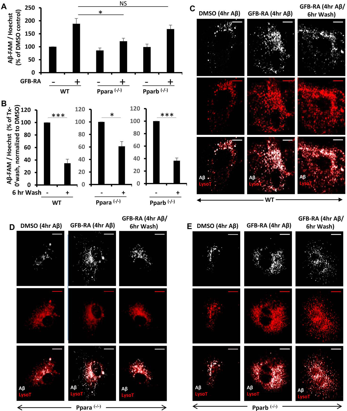
Figure 3. Role of PPARα and PPARβ in Aβ uptake and degradation in mouse primary astrocytes.

(A) Mouse primary astrocytes isolated from WT, Ppara−/− and Pparb−/− animals were treated with GFB-RA [GFB (10 μM) + RA (0.2 μM)] or DMSO, followed by incubation with 500 nM FAM-Aβ and subjected to Aβ uptake assay. Data were compared to DMSO-treated WT control and represented as percent change. (B) Mouse primary astrocytes isolated from WT, Ppara−/− and Pparb−/− animals were, treated with GFB-RA, followed by incubation with 500 nM FAM-Aβ for 4 hours, washed in Aβ free media for 6 hours, and subjected to an Aβ degradation assay. All data are representative of the mean ± SEM of three independent experiments run in duplicate. *P<0.05, ***P<0.001, and NS denotes not significant by one-way ANOVA followed by Dunnett’s multiple comparison test. (C to E) Mouse primary astrocytes isolated from WT (C), Ppara−/− (D) and Pparb−/− (E) animals were isolated, treated with DMSO, followed by incubation with 500 nM HF-647-Aβ for 4 hours and 75 nM Lysotracker for 45 min (first panel), treated with GFB-RA, followed by incubation with 500 nM HF-647-Aβ for 4 hours and 75 nM Lysotracker for 45 min (second panel), treated with GFB-RA, followed by incubation with 500 nM HF-647-Aβ for 4 hours and washed in Aβ-free media for 6 hours and incubated in 75 nM Lysotracker for 45 min (third panel) and observed under microscope. Scale bar = 20 μm. Results are representative of three independent experiments run in duplicate.
