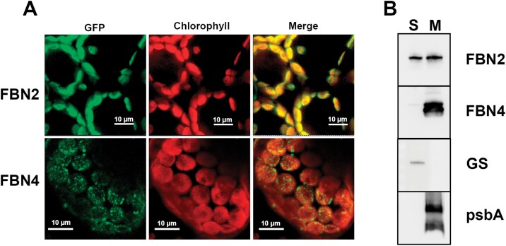
Fig. 2.
Localization of FBN2. (A) Full-length FBN2 or FBN4 cDNAs were fused to GFP and transiently expressed in N. benthamiana leaves. Fluorescence was monitored by confocal microscopy. The GFP fluorescence, chlorophyll autofluorescence, and merged images are shown. (B) Chloroplasts isolated from Arabidopsis rosette leaves were disrupted and the soluble (S) and membrane fractions (M) were isolated by ultracentrifugation at 100000 g for 1h at 4 °C. The pellet (membrane fraction) was resuspended in the same volume as the supernatant and 20 µg of protein from each fraction was loaded on to SDS-PAGE gels. The proteins were blotted on to a PVDF filter and hybridized with specific antibodies against FBN2, FBN4, plastidial glutamine synthetase (GS) (a marker of stromal protein), and psbA (a marker of thylakoid membranes).

