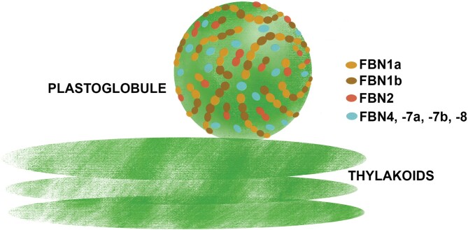
Fig. 8.
Schematic model of the arrangement of FBNs1-2 subgroup proteins on the surface of PGs. FBN2 (red) may form homodimers or heterodimers with FBN1a (light brown) or FBN1b (dark brown). We have previously shown that FBN1a and FBN1b may form hetero-oligomers (Gámez-Arjona et al, 2014a). These interactions allow the formation of a FBNs1-2-based network around the surface of PGs. Other proteins, such as those described in Table 1, associate with PGs via interactions with these FBNs. The degree of functional redundancy between these FBNs has not been characterized and might vary for each PG-associated protein. Their elimination would affect the localization and function of some PG-associated proteins. The functions of other FBNs associated with PGs (FBN4, FBN7a, FBN7b, and FBN8, indicated in light blue) have not been determined yet.

