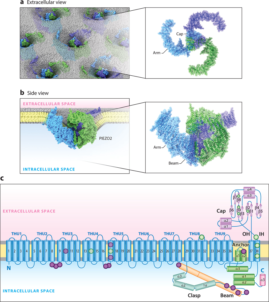
Figure 1.
The structure of mouse PIEZO2. (a) A top-down illustration of mouse PIEZO2 channels in the membrane as viewed from outside the cell. (b) A side-view illustration of a PIEZO2 channel curving the plasma membrane. (c) Ribbon diagram of one blade of PIEZO2 highlighting key functional domains. Lavender and green circles indicate approximate reported locations of human loss-of-function and gain-of-function variants, respectively. Structure adapted with permission from Reference 48. Abbreviations: α, α-helix; β, β-pleated sheet; IH, inner helix of the ion-conducting pore; OH, outer helix of the ion-conducting pore; THU, transmembrane helical unit.

