Figure 6.
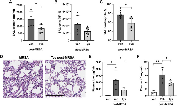
Effects of posttreatment with Tys on MRSA-induced acute lung injury. Wild-type mice were treated with live bacteria-MRSA (0.75 × 10⁸ CFU/mouse, it) or PBS (control), and 1 h later received Tys (0.5 mg/kg, ip). BAL protein (A), total cell counts (B), and neutrophil percentages were determined (C). D: H&E lung staining. Lung tissues were scanned with a digital slide scanner at ×40 and representative pictures were taken at ×20. Scale bar: 200 μm. Representative images are shown. IL-6 (E) and KC (F) levels in plasma. n = 3–6 mice per condition. Unpaired t test (A–C), one-way ANOVA (E and F), *P < 0.05, **P < 0.01. BAL, bronchoalveolar lavage; CFU, colony-forming unit; H&E, hematoxylin and eosin; MRSA, methicillin-resistant Staphylococcus aureus; PBS, phosphate-buffered solution.

