Figure 7.
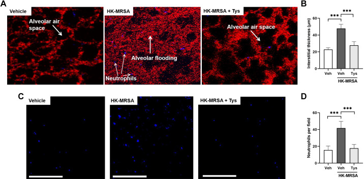
Intravital microscopy demonstrates attenuation of MRSA-induced neutrophil recruitment and interstitial edema after treatment with Tys. Wild-type mice were treated with Tys (0.5 mg/kg, ip) or vehicle 1 h before administration of HK-MRSA (2 × 10⁸ CFU/mouse, it) or PBS (control). Live animal imaging by two-photon microscopy was performed 18 h following treatment. A: representative images showing the interstitial space visualized by TMR-labeled dextran (red) between alveolar airspaces (black) in each condition. Individual neutrophils labeled with fluorescent Gr1 antibody appear purple. B: quantification of interstitial thickness in µm. Additional representative images showing only fluorescent-labeled neutrophils (purple dots; C), and quantification of neutrophils per field (D). White scale bar = 200 μm, n = 5 mice per group. One-way ANOVA, ***P < 0.001. CFU, colony-forming unit; HK-MRSA, heat-killed MRSA; MRSA, methicillin-resistant Staphylococcus aureus; PBS, phosphate-buffered solution; TMR, tetramethylrhodamine.

