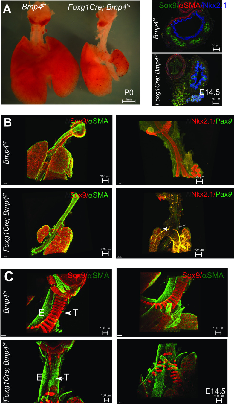
Figure 2.
Mesenchymal deletion of Bmp4 impairs respiratory tract development and patterning of the tracheal mesenchyme. A: postnatal day (P)0 image of Foxg1Cre; Bmp4f/f respiratory tract depicts the poorly developed trachea and lungs compared with the control. Cross section staining of embryonic day (E)14.5 trachea shows that deletion of Bmp4 from the mesenchyme renders poor development with nonexistent cartilage on the ventral side, resulting in a flaccid trachea. Respiratory identity was confirmed by NKX2.1 (blue) staining. B: whole mount immunofluorescence of E14.5 Foxg1Cre;Bmp4f/f truncated tracheas stained for smooth muscle [α-smooth muscle actin (αSMA), green] and cartilage (SOX9, red) reveal the ectopic presence of muscle on the ventral side and mispatterned or reduced cartilage near the carina and bronchi. Trachea and esophagus were stained with NKX2.1 (red) and PAX9 (green), respectively. In Foxg1Cre;Bmp4f/f, the carina and bronchi (arrowheads) are adjacent to the esophagus (arrow) and seem to emerge from a blind tube as opposed to the continuous structure in the control. C: whole mount immunofluorescence of E14.5 Foxg1Cre;Bmp4f/f “complete” tracheas revealed nearly nonexistent cartilage (SOX9, red) in the trachea with poor mesenchymal condensations in carina and bronchi and anomalous presence of smooth muscle (αSMA, green) in the ventral aspect of the tracheal mesenchyme. E, esophagus; T, trachea.

