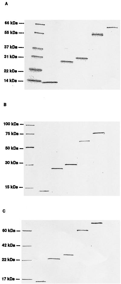
FIG. 1.
Characterization of expressed recombinant proteins. The separation of proteins by SDS-PAGE was performed in three identical gels, one of which was stained for protein visualization with Coomassie blue (A). The two other gels were transferred to a Hybond C-extra nitrocellulose membrane and immunodetected with either anti-His antibody (penta-His antibody) according to Qiagen instructions for 6xHis-tagged protein detection (B) or a pool of 22 sera from patients with positive urethral or endocervical C. trachomatis DNA amplification for IgG-specific antibody binding (C). Lanes (left to right) MW standard, hsp10, MIP, pgp3, hsp60, and hsp70.

