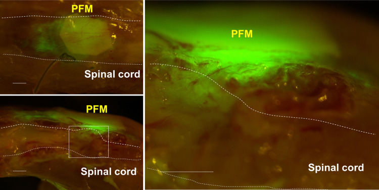
Fig 3. Expansion of implanted HAP stem cells in the severed spinal cord.
GFP-expressing HAP stem cells migrated out from the PFM and joined the severed thoracic spinal cord. Left panel = low-magnification of coronal section (upper) and sagittal section (lower). Right panel = high-magnification of white boxed area. Bar = 500 μm.

