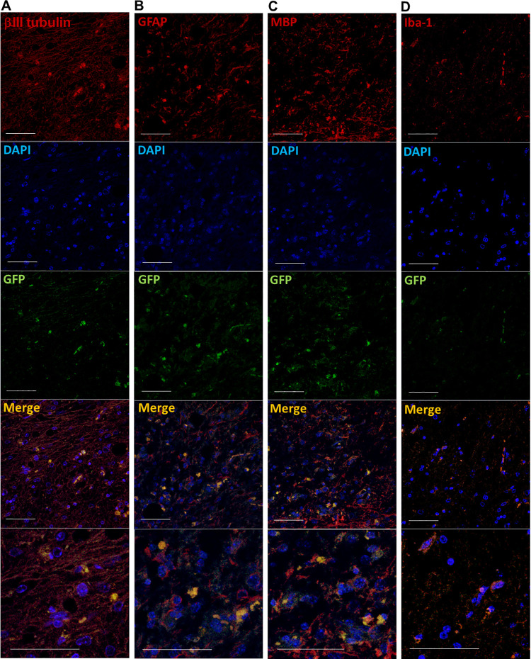
Fig 4. Immunofluorescence staining of the severed spinal cord implanted with HAP stem cells.
Immunofluorescence staining shows that in the joined area of the previously severed spinal cord, the implanted HAP stem cells differentiated to neurons (A), astrocytes (B), oligodendrocytes (C). Microglia were GFP negative in the joined area of severed spinal cord (D). Red = βIII tubulin (A), GFAP (B), MBP (C) or Iba-1 (D); Green = GFP (A-D); Blue = DAPI (A-D); Merged (A-D). Bar = 50 μm. All images show sagittal sections of the spinal cord.

