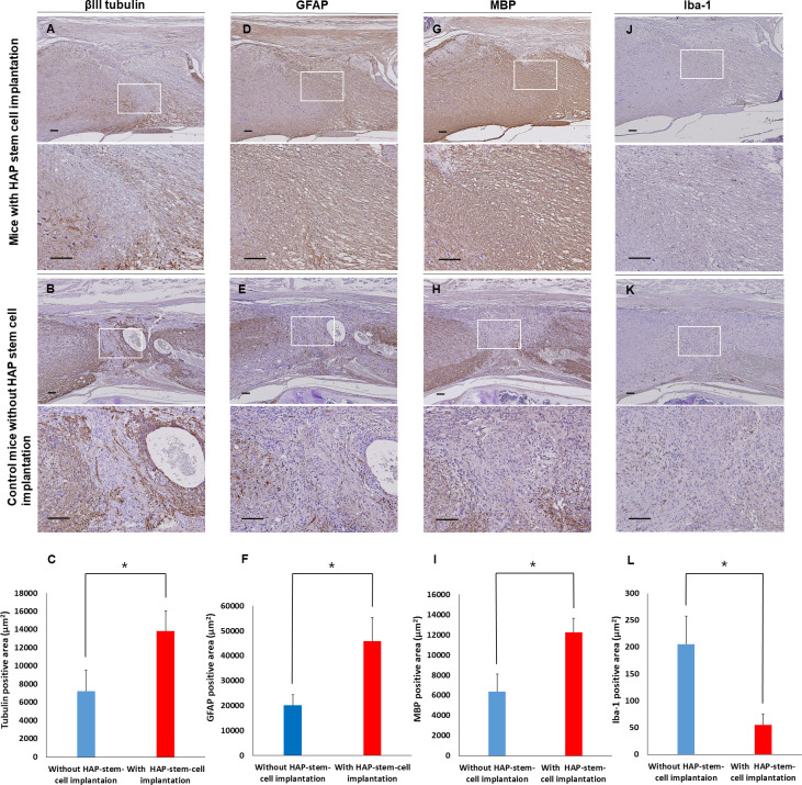
Fig 5. Immunostaining of the severed spinal cord implanted with HAP stem cells.
(A, B) βIII tubulin expression in the repaired spinal cord after HAP-stem-cell implantation and without implantation. (C) The βIII tubulin-positive area in sagittal sections in mice with implanted HAP stem cells and control mice (mice with HAP-stem-cell implantation: n = 5; control mice without implantation: n = 5). *P < 0.05. (D, E) GFAP expression in the repaired spinal cord after HAP stem cell implantation and without implantation. (F) The GFAP-positive area in sagittal sections in the mice with implanted HAP stem cells and control mice (mice with HAP-stem-cell implantation: n = 5; control mice without implantation: n = 5). *P < 0.05. (G, H) MBP expression in the repaired spinal cord after HAP-stem-cell implantation and in the un-repaired spinal cord without implantation. (I) The MBP-positive area in sagittal sections in the mice with implanted HAP stem cells and control mice with an un-repaired spinal cord (mice with HAP-stem-cell implantation: n = 5; control mice without implantation: n = 5). *P < 0.05. (J, K) Iba-1 expression in the repaired spinal cord after HAP-stem-cell implantation and in the un-repaired spinal cord without implantation. (F) The Iba-1-positive area in sagittal sections in the mice with implanted HAP stem cells and control mice without HAP-stem-cell implantation (mice with HAP-stem-cell implantation: n = 5; control mice without implantation: n = 5). *P < 0.05. Upper panels = low-magnification. Bar = 100 μm. Lower panels = high-magnification of white boxed area. Bar = 100 μm. All images show sagittal sections with the spinal cord.

