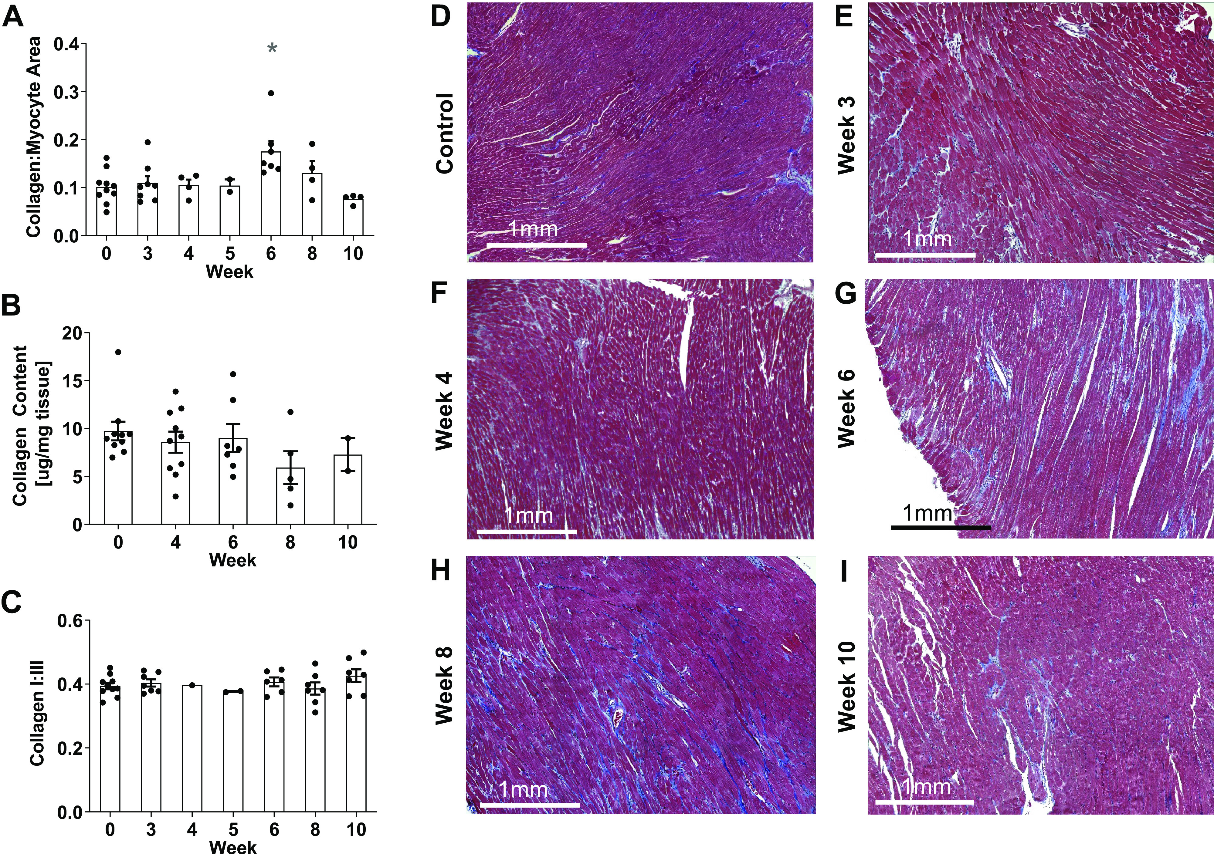
Figure 6.

Histological collagen to myocyte area fraction (A) from Masson’s trichrome-stained RV sections in control (D) rats and SuHx rats at weeks 3 (E), 4 (F), 6 (G), 8 (H), and 10 (I) was only significantly different from control at week 6 (*P < 0.05). RV myocardial total collagen content (B) and collagen type I to III ratio (C) showed no significant changes throughout the study (P > 0.05). Data are shown as means ± SE compared with the control group. RV, right ventricle; SuHx, sugen-hypoxia.
