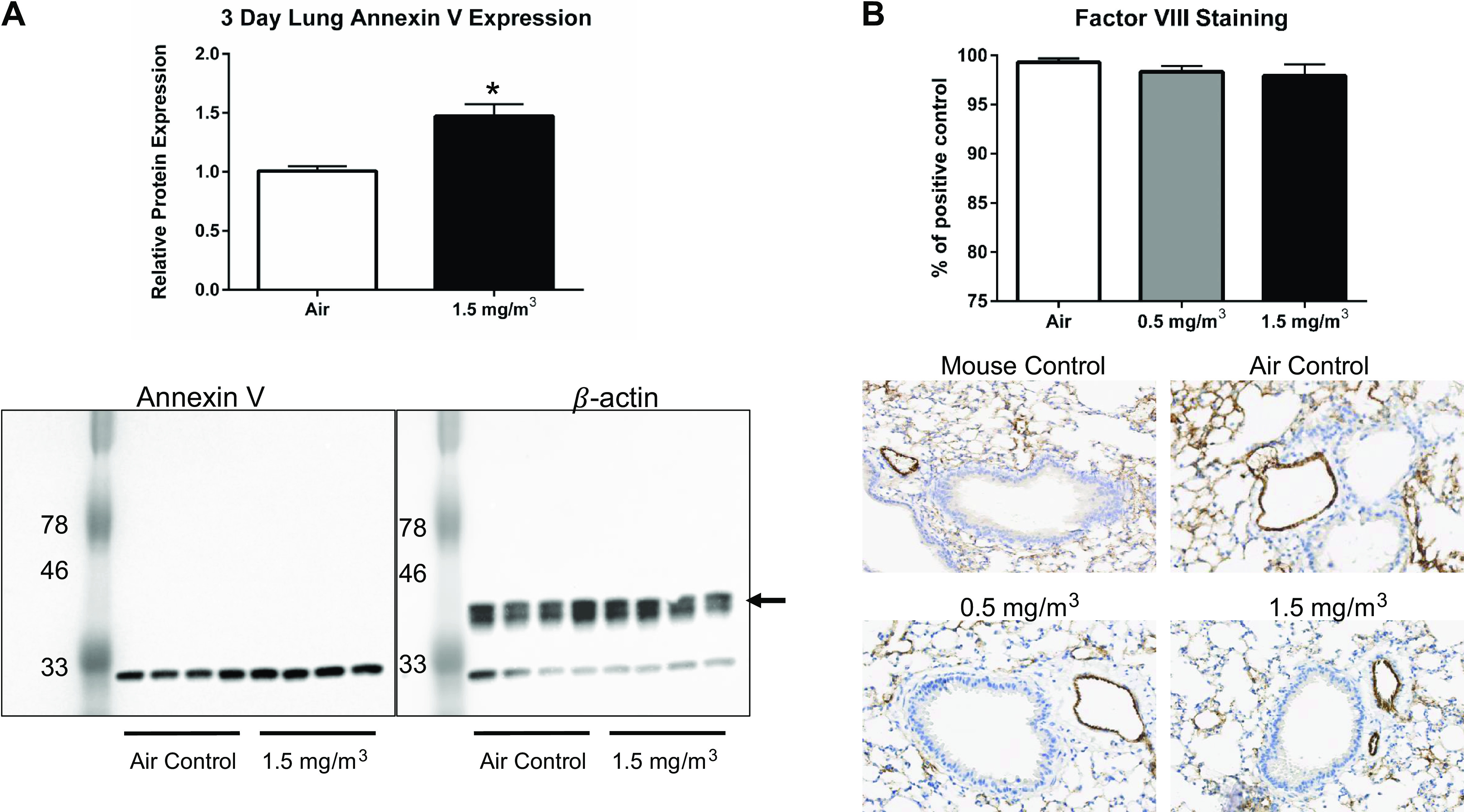
Figure 9.

DCB230 exposure increased apoptosis marker Annexin V in the lung, but did not disrupt the integrity of the vascular endothelium. A: Annexin V protein levels were measured in lung tissue homogenates by Western blot analysis. The blot was probed first for Annexin V protein (left) and then for β-actin (right) as a housekeeping protein. Note the lower band in the right panel represents residual Annexin V staining. *P < 0.05. B: the endothelium of lung cross sections was visualized by immunohistochemical staining for factor VIII protein. Representative sections are shown for control mouse lung compared with the lungs of mice exposed to filtered air, 0.5 mg/m3 or 1.5 mg/m3 DCB230. Staining in images were quantified using Image J software and the data are expressed as a % of the control (normal mouse lung).
