Figure 1.
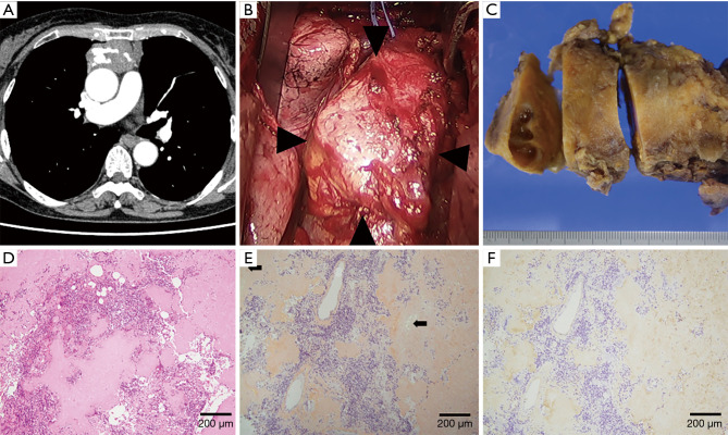
Thymic amyloidosis (Case 1). (A) Chest CT showed a solid, large mass with calcification located in the anterior mediastinum in the mediastinal window. (B) A hard tumor with a smooth surface originated from the thymus gland and firmly adhered to the pericardium alone (arrowheads). The left innominate vein was encircled using vessel tape. (C) The resected tumor measured 100 mm in maximum diameter, had a smooth appearance, and was extremely firm. The cut surface was light yellow with cystic lesions. (D) The tumor included diffuse deposits of amorphous and eosinophilic material, calcification, lymphocytes and plasma cells, but no neoplastic cells (hematoxylin and eosin stain). (E) Amorphous materials were positive in direct fast scarlet 4BS staining and showed an “apple-green” birefringence under polarized light (arrows). (F) Immunohistochemical staining was positive for κ light chain.

