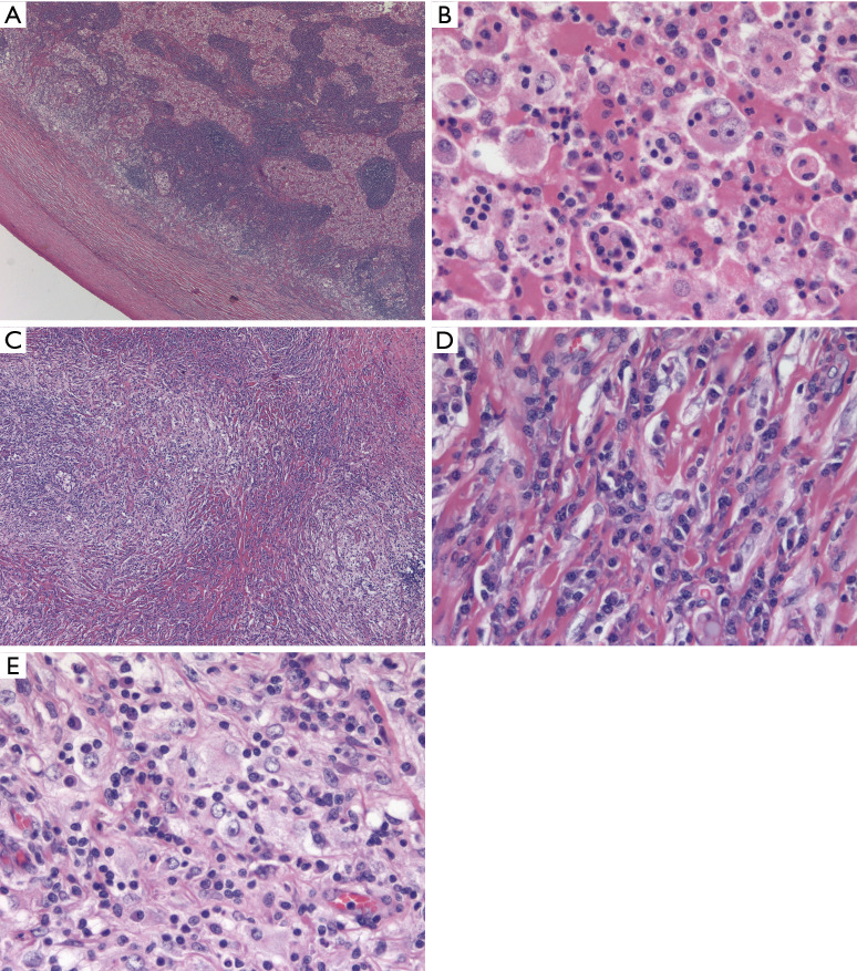
Figure 1.
RDD. (A) Classic low power appearance of nodal RDD with a thickened capsule and a serpiginous anastomosing network of expanded sinuses distended by large histiocytes with abundant eosinophilic cytoplasm (H&E stain, 25× magnification). (B) Higher magnification (400×) shows prototypical emperipolesis, with small inflammatory cells appearing superimposed upon the cytoplasm of voluminous histiocytes with large vesicular nuclei and prominent central nucleoli. (C) Same case with transition to distinct morphology of extranodal RDD with areas of marked sclerosis alternating with more cellular, paler areas (H&E stain, 100× magnification). (D) Typical sclerotic areas of extranodal RDD consist of large numbers of plasma cells with rare interspersed RDD histiocytes, which could easily be overlooked on a small biopsy (H&E stain, 400× magnification). (E) Looser areas of extranodal RDD with dense lymphoplasmacytic infiltrate with interspersed RDD histiocytes (H&E stain, 400× magnification). RDD, Rosai-Dorfman disease.

