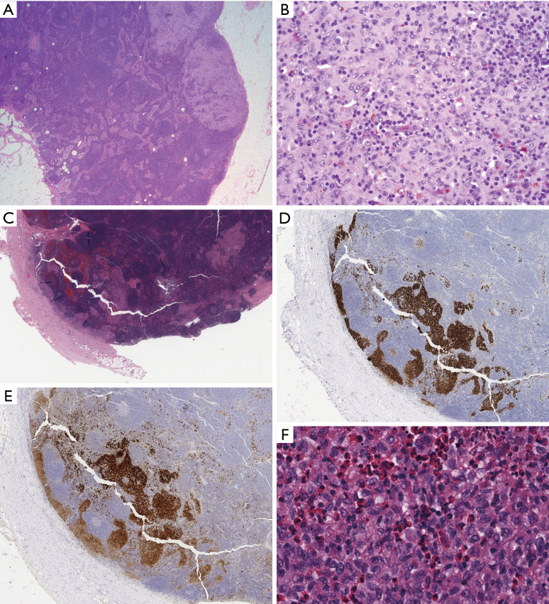
Figure 2.
Nodular paracortical hyperplasia versus nodal involvement by LCH. (A) Typical low power appearance of nodular paracortical hyperplasia, showing pale nodular aggregates within lymph node parenchyma (H&E stain, 25× magnification); (B) higher power of paracortex shows numerous Langerhans cells with abundant eosinophilic cytoplasm, and angulated, vesicular nuclei admixed with numerous small lymphocytes (H&E stain, 400× magnification); (C) in contrast, at low power, nodal involvement by LCH replaces the subcapsular sinus (H&E stain, 50× magnification). The sinus distribution is highlighted by immunohistochemistry for CD1a (D, 50× magnification) and S100 (E, 50× magnification), which also highlights scattered and singly distributed interdigitating dendritic cells. (F) High magnification demonstrates similar cytologic features including nuclear angulation and grooves, but numerous admixed eosinophils are indicative of LCH (H&E stain, 400× magnification). LCH, Langerhans cell histiocytosis.

