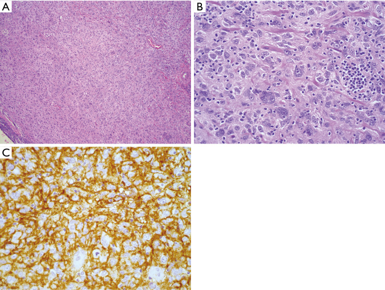
Figure 3.
FDCS. (A) H&E stained section (100×) showing a histiocytoid cell proliferation with a loose whirling architecture; (B) higher power (400×) demonstrates tumor cells, many with eosinophilic nucleoli, and prominent admixed small, mature lymphoid cells; (C) strong CD35 expression confirming follicular dendritic cell lineage (immunohistochemistry, 400× magnification). FDCS, follicular dendritic cell sarcoma.

