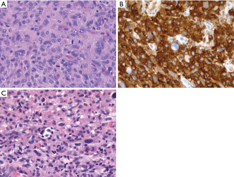
Figure 4.
HS. (A) Syncytial sheets of markedly atypical cells with eosinophilic cytoplasm, prominent nucleoli, and mitotic activity (H&E stain, 400× magnification); (B) diffuse CD163 reactivity (immunohistochemistry, 400× magnification); (C) separate case showing sheets of tumor cells with marked nuclear pleomorphism, foamy cytoplasm, and scattered admixed non-neoplastic inflammatory cells (H&E stain, 400× magnification). The morphology of histiocytic sarcoma is not specific and would be compatible with other pleomorphic tumors given a different immunoprofile. Thus, diagnosis of HS requires a broad panel of immunohistochemical stains to exclude other tumors. HS, histiocytic sarcoma.

