Figure 6.
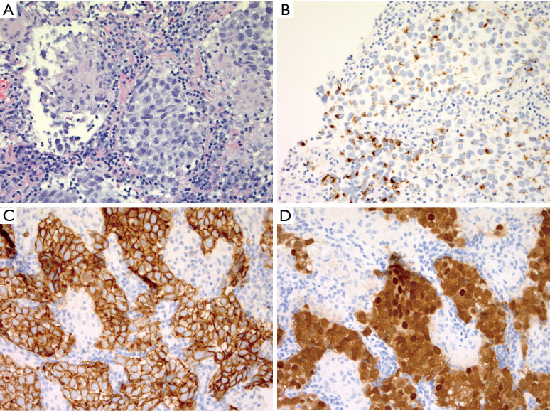
Mediastinal germinoma with histiocytic infiltrate. (A) Core biopsy showing high magnification comparison between histiocytic and giant cell reaction (left) versus sheets of large epithelioid germinoma cells with vesicular nuclei (H&E, 400× magnification). (B) Importantly, focal keratin expression can be seen in germinoma (immunohistochemistry, 400× magnification), and if considered alongside diffuse CD117 expression (C, 400× magnification), this lesion could easily be mistaken for thymic carcinoma. The nested architecture and sclerosis would certainly also raise the differential diagnosis of primary mediastinal large B-cell lymphoma. (D) Nuclear OCT3/4 and SALL4 expression (not pictured) are confirmatory (immunohistochemistry, 400× magnification).

