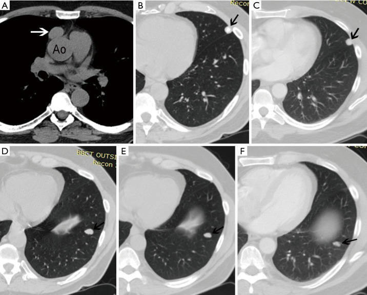
Figure 1.
A 58-year-old man with newly diagnosed prevascular mediastinal mass. Unenhanced chest CT demonstrates a spherical 2.7×1.8 cm ovoid well-demarcated soft tissue mass (arrow in A) in the prevascular mediastinum, abutting the ascending aorta (Ao) but separated from it by a sliver of fat. The pleura is normal, but there are two pulmonary nodules. One is benign as it is heavily calcified (arrow in B) and remained stable compared to a CT performed 9 years earlier (arrow in C). The other nodule, in the left lobe (arrow in D), is lobulated, solid left, and not calcified. It gradually grew over the years currently measuring 1.6×1.1 cm in diameter, whereas in a preceding CT performed 4 years earlier it measured 1.4×0.8 cm in diameter (arrow in E), and 9 years prior to the current CT it measured 1.2×0.7 cm in diameter (arrow in F).

