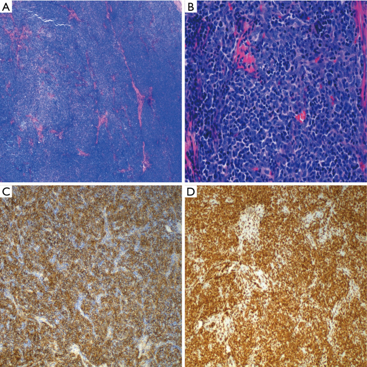
Figure 2.
(A) A low power view shows a lobulated neoplasm. The hypercellular lobules are focally intersected by fibrous band; (B) at high power, the cellular lobules are comprised predominantly of small lymphocytes. Scattered larger epithelioid cells (neoplastic cells) are also identified; (C) a keratin stain highlights a meshwork of keratin-positive neoplastic cells; (D) the Tdt stain highlights the thymocytes.

