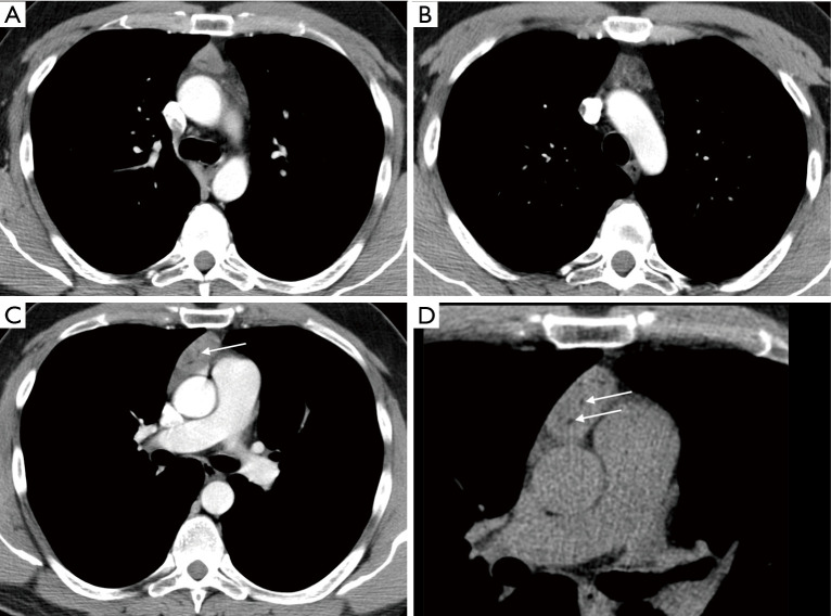
Figure 1.
Computed tomography (CT) of the mediastinal tumor. The axial images of contrast enhanced chest CT showed the slightly enlarged thymus (A) with rounded contours and a solid mass within it (B). The mass was discreetly heterogeneous with some small fatty bands (C - arrow). Infiltration of vessels and tumor calcifications were not demonstrated (D).

