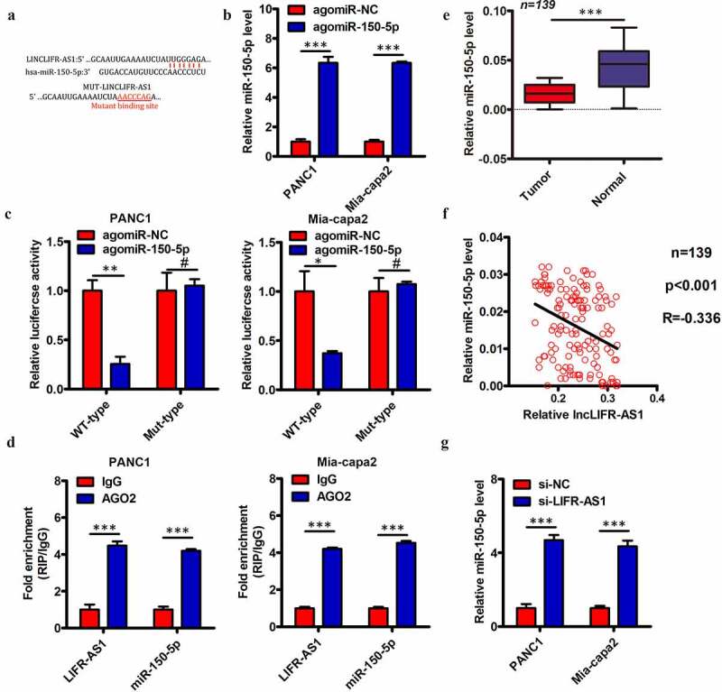
Figure 4.

LIFR-AS1 interacts with and sequesters miR-150-5p within PC cells
(a) miR-150-5p interaction with LIFR-AS1 was predicted via bioinformatic analyses. (b) The expression of miR-150-5p was quantified in PANC1 and Mia-capa2 cells that had undergone agomir-150-5p or agomir-NC transfection. (c) Wild type (WT)-LIFR-AS1 or mutant (MUT)-LIFR-AS1 were co-transfected into PANC1 and Mia-capa2 cells along with agomir-150-5p or agomir-NC, and after 48 h luciferase activity was measured. (d) Higher levels of LIFR-AS1 and miR-150-5p were detectable in immunoprecipitates containing Ago2 relative to those prepared using a control IgG. (e) Levels of miR-150-5p expression in 139 pairs of PC and paracancerous control tissues were quantified via qRT-PCR. (f) A Spearman’s correlation analysis of the relationship between miR-150-5p and LIFR-AS1 expression in PC tissue samples from E was conducted. (g) Levels of miR-150-5p were measured via qRT-PCR in PANC1 and Mia-capa2 cells in which LIFR-AS1 had been knocked down. *P < 0.05; **P < 0.01; ***P < 0.001; #P > 0.05.
