Figure 3.

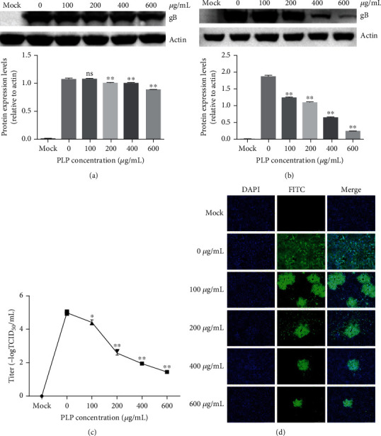
PLP inhibits PRV attachment and penetration into PK15 cells. (a) We treated PK15 cells by PLP at diverse doses for a 1 h period and then infected by PRV XJ5 (MOI = 0.1) with PLP. At 24 hpi, we conducted WB assay for evaluating β-actin and PRV gB protein levels. (b) We infected PK15 cells by PRV XJ5 (MOI = 0.1) for a 1 h period under 4°C with PLP at diverse doses and incubated with different concentrations of PLP at 37°C for a 1 h period prior to removal of PLP DMEM as well as cultivated within PLP-free DMEM. Protein levels of β-actin and PRV gB were measured through WB at 24 hpi. (c) We determined virus titers through TCID50. (d) We assessed internalized virus through IFA. The error bars indicated SD of 3 individual assays. ∗P < 0.05, ∗∗P < 0.01.
