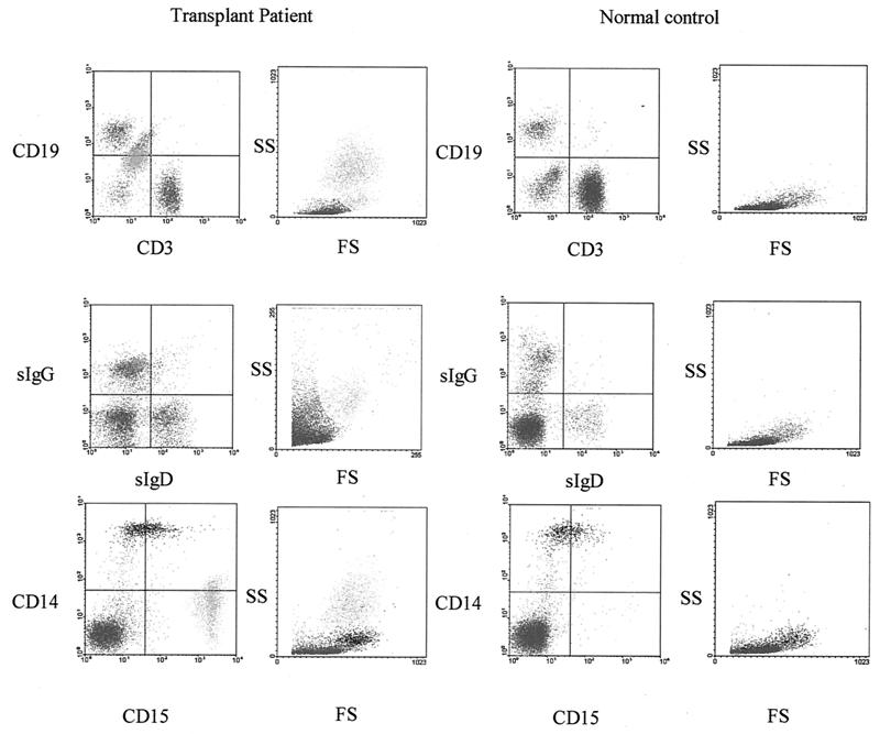
FIG. 2.
Characterization of the CD19hi and CD19lo cell populations detected in the peripheral blood lymphocyte fraction. (Top row) Flow cytometric analysis of B-cell (CD19) and T-cell (CD3) markers after gating on the population of high-FS and -SS cells unique to lymphocyte preparations of transplant recipients compared to a normal healthy EBV-positive control donor. (Center panels) Analysis of the sIgG and sIgD expression. (Bottom panels) Analysis of monocyte (CD14) and granulocyte (CD15) markers. High-FS and -SS cells appear to be CD19lo, IgG+, and CD15+.

