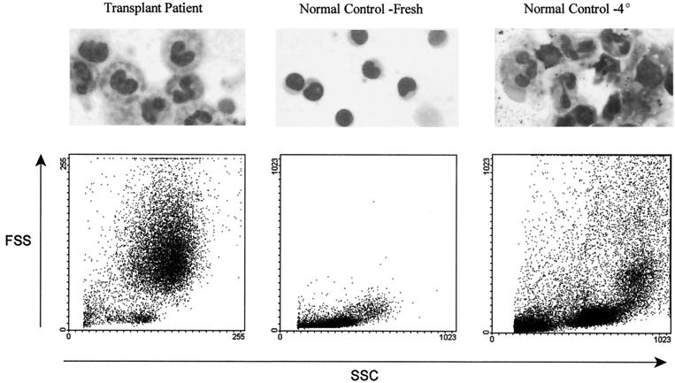
FIG. 3.
Wright staining of lymphocyte preparations from a pediatric transplant recipient (left panel) reveals the presence of polymorphonuclear cells, and the light-scattering profile contains large numbers of high-FS and -SS cells. A normal control donor lymphocyte preparation (center panel) has no polymorphonuclear cells and no high-FS and -SS cells. Incubation of a blood specimen at 4°C overnight prior to Ficoll-Hypaque gradient preparation shows the appearance of polymorphonuclear cells and high-FS and -SS cells (right panel)

