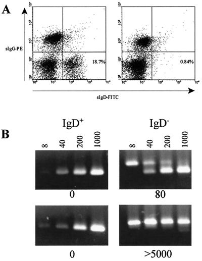
FIG. 5.
EBV viral load in naive B cells fractionated from lymphocyte preparations from chronic viral load carriers. (A) Flow cytometric profiles of sIgG- and sIgD-stained cells before (left panel) and after (right panel) anti-IgD magnetic bead separation of the naive B cells. (B) Ethidium bromide-stained DNA products of QC-PCRs using 104 IgD+ or 105 IgD− cells and 8, 40, 200, or 1,000 copies of an identical competitor target sequence were analyzed by agarose gel electrophoresis. Results for patient 7, a low-load carrier, and patient 15 (a high-load carrier) are shown. The interpolated DNA concentration in copy numbers of EBV genomes detected is shown below each panel. A complete analysis of all 19 patients in the study is summarized in Table 1.

