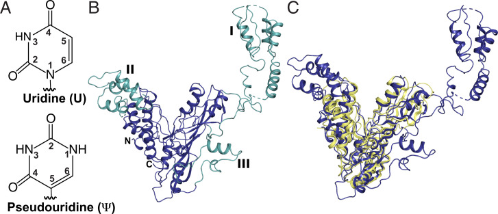
Fig. 1.
S. cerevisiae Pus7 structure. (A) Uridine and pseudouridine. (B) X-ray structure of Pus7 at 3.2-Å resolution (PDB: 7MZV). The structurally conserved, V-shaped enzyme core housing the PUS and TRUD domains (blue). The three eukaryotic-specific insertions (green) are numbered I through III. (C) Superimposition of the S. cerevisiae Pus7 (blue) and E. coli TruD (yellow, PDB: 1SB7) structures demonstrating the structural conservation of the enzyme’s catalytic core.

