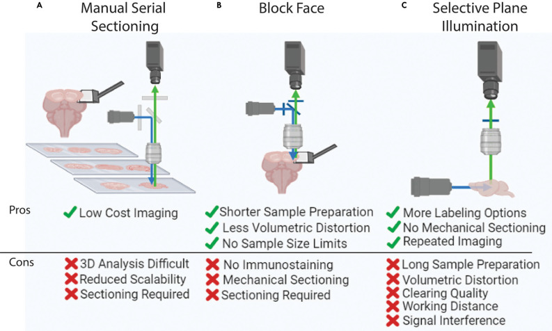
Figure 3.
Whole brain imaging methods. (A) Manual cutting and imaging histological sections on slides. This method is laborious and requires significant amounts of manual effort. (B) Block-face imaging such as serial two photon tomography. The requirement of serial sectioning can introduce challenges in delicate samples. (C) Selective plane illumination or light-sheet imaging. This method can visualize an intact and clear sample without any physical sectioning, yet this imaging is hard to apply to large tissue (e.g., human brain) due to the limited working distance of an objective lens.

