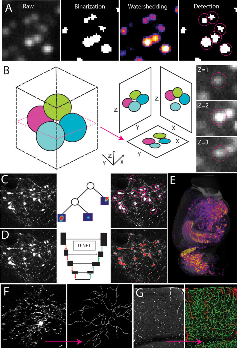
Figure 4.
Automated signal detection. (A) Cell detection pipeline using filter-based methods including intensity filters and water-shedding. (B) Cells can be captured in multiple planes making it necessary to identify the connected components of a central point in the sphere. (C–E) Random forest-based machine learning (C) or deep learning segmentation (D) to detect cell type signals across the whole brain (E). (F,G) Signal detection and tracing to achieve full 3D reconstruction of a single neuron (F, adapted from Janelia MouseLight NeuronBrowser) or cerebrovascular network analysis (G).

