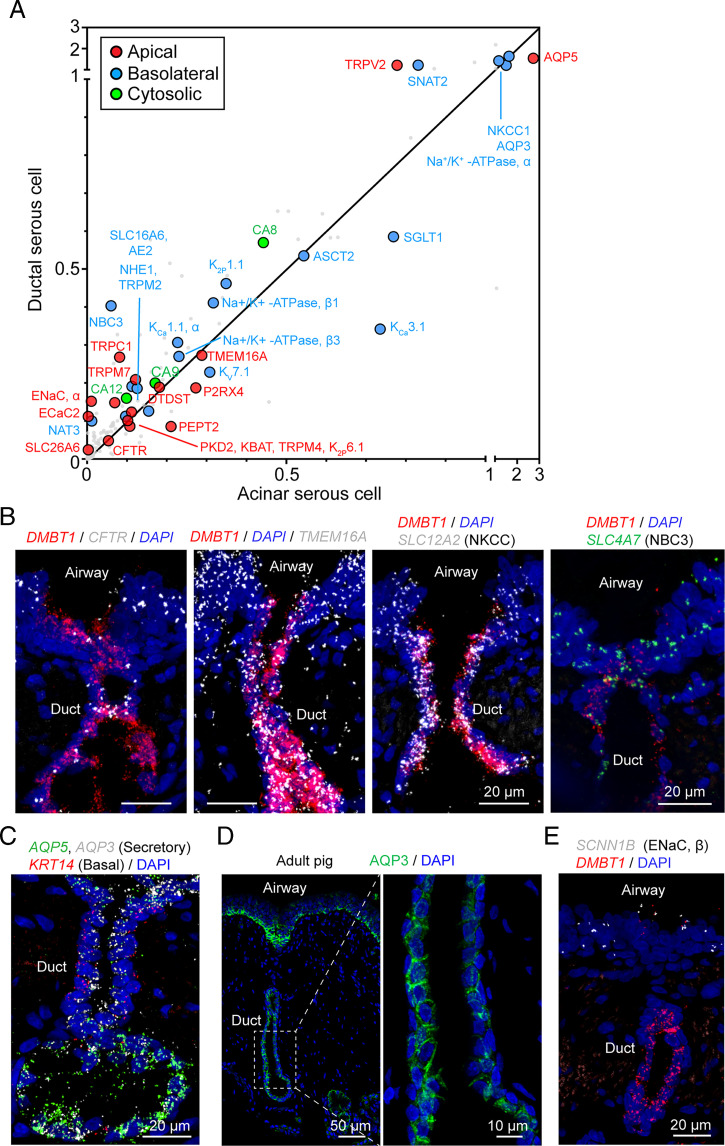
Fig. 7.
SMG ductal serous cell transcripts indicate an anion secretory function. (A) Scatter plot of the mean expression of transepithelial electrolyte transporter genes in ductal and acinar serous cells at natural log scale. Different colors of dots indicate different subcellular localization of the encoded protein. (B) Representative images of the expression pattern of key ion transporters (CFTR, TMEM16A, NKCC, NBC3) in the duct of SMGs. Ductal serous cells (DMBT1+) are shown in red. (C) Representative image of smFISH of AQP3, AQP5, and KRT14 in tracheal sections. In SMGs, AQP3 and AQP5 are expressed in both ductal serous cells and acinar serous cells. KRT14 is expressed in basal cells of the duct and myoepithelial cells of the acinus. (D) Representative images of immunostaining of AQP3 in airway sections of an adult pig. Enlarged image shows that AQP3 is localized at the basolateral region of ductal serous cells. (E) Representative images of smFISH of SCNN1B (encodes the β-subunit of ENaC) in tracheal sections. SMG duct (DMBT1+ region, red) was not labeled with SCNN1B signal.

