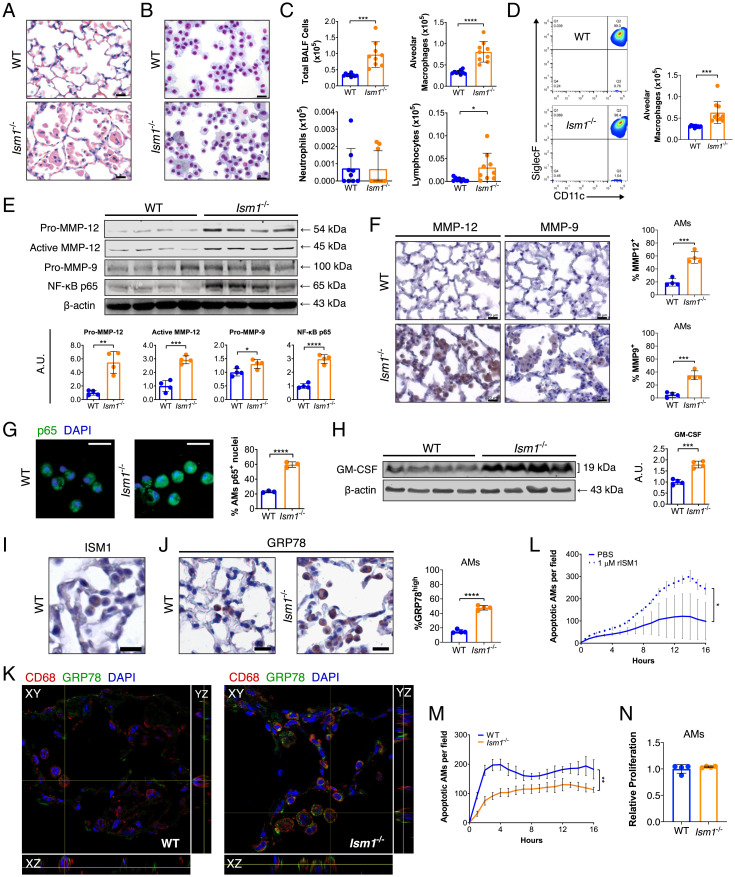
Fig. 2.
Ism1−/− mouse lungs present up-regulated COPD mediators at 2 mo of age. (A) H&E stained lungs showing focal AM accumulation in FVB/NTac Ism1−/− mice. (B) Liu-stained cytospins and (C) quantifications of BALF cells from FVB/NTac WT and Ism1−/− lungs. (D) Flow cytometric analysis and quantifications of AMs (Siglec F+ CD11c+) in BALF from FVB/NTac WT and Ism1−/− lungs. (E) Western blots and quantification of fold changes for Pro-MMP-12, Active-MMP-12, Pro-MMP-9, and NF-κB p65 with β-actin as loading control in FVB/NTac WT and Ism1−/− lungs. (F) IHC and quantifications of MMP-12+ and MMP-9+ AMs of FVB/NTac WT and Ism1−/− lungs. (G) IF staining for NF-κB p65 with nuclei counterstain (DAPI) and quantification of primary AMs harboring nuclear p65+ from FVB/NTac WT and Ism1−/− mice. (H) Western blot and fold change for GM–CSF with β-actin as loading control in FVB/NTac WT and Ism1−/− lungs. (I) IHC for ISM1, and (J) IHC for GRP78 and quantification of GRP78high AMs in FVB/NTac WT and Ism1−/− mice. (K) Confocal fluorescent microscopy image demonstrating csGRP78high AMs in FVB/NTac WT and Ism1−/− lungs. (L) rISM1 induces apoptosis in WT primary AMs. (M) Apoptosis of freshly isolated primary AMs from FVB/NTac WT and Ism1−/− lungs. Analysis was carried out in triplicate or quadruplicate wells. (N) Proliferation of primary AMs from WT and Ism1−/− mice. Analysis was carried out in triplicate wells. Data are mean ± SD and were analyzed by two-group, two-tailed Student’s t test (C–H, J, and L–N). *P < 0.05, **P < 0.01, ***P < 0.001, and ****P < 0.0001. n = 3 to 10 mice per group. A.U.: arbitrary units. (Scale bars, 20 μm for A, B, F, G, I, and J.) Data from C and D are integrated from two independent experiments using different WT and Ism1−/− mice (biological repeats, n = 9 to 10). Data from E–M are representatives of twice-repeated experiments with similar results. Data from N is from one independent experiment.

