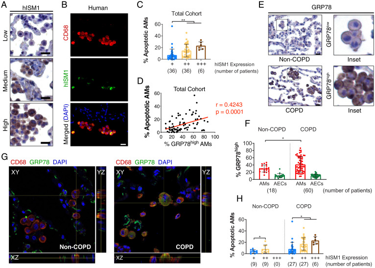
Fig. 4.
hISM1 expression correlates with AM apoptosis. (A) IHC staining for hISM1 and (B) double IF staining for CD68 and hISM1 with nuclei (DAPI) counterstain in lung tissue sections showing AM expression of hISM1. (C) Quantifications of AM apoptosis in different hISM1 expression level groups. (D) Correlation analyses between high GRP78 expression with AM apoptosis. (E) Representative IHC for GRP78 depicting low GRP78 expression in non-COPD AMs and high GRP78 expression in COPD AMs. (F) Percentage of AMs and alveolar epithelial cells (AECs) in non-COPD and COPD patients with GRP78high expression (E, COPD Inset). (G) Confocal microscopy images for CD68, GRP78, and nuclei (DAPI) in non-COPD and COPD human lungs. (H) Percentage of apoptotic AMs stratified by COPD status and hISM1 expression. Data are mean ± SD and were analyzed by Pearson correlation (D), one-way ANOVA (C), and two-way ANOVA (F and H) with Tukey’s post hoc test. Patient sample sizes are depicted on graphs. (Scale bars, 20 μm for A, B, and E.)

