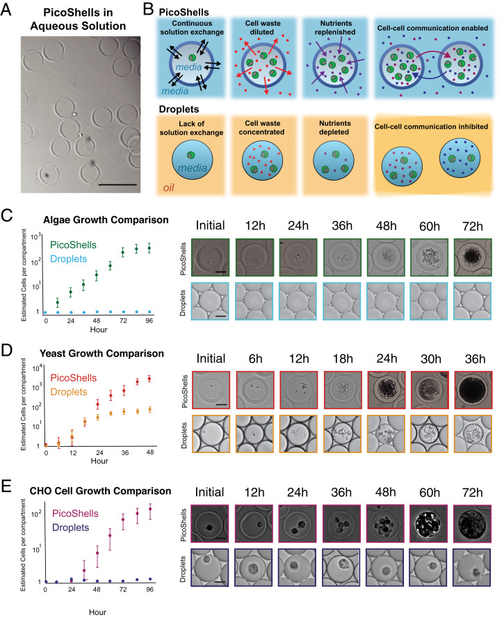
Fig. 2.
Growth comparison between PicoShells and emulsion droplets. (A) PicoShells are solid spherical particles that contain a hollow inner cavity and a porous outer shell. (Scale bar: 200 µm.) (B) PicoShells allow for continuous solution exchange with the external environment such that cell waste can be diluted, nutrients can be replenished, and cell–cell communication factors can pass between adjacent PicoShells. (C) Chlorella were encapsulated into PicoShells and droplets to compare division rates in each compartment. Results show that the microalgae do not grow in droplets but grow readily in the particles; 5,000 to 6,000 cell–containing PicoShells/droplets were analyzed for each time point. Error bars represent the SD in the estimated number of cells per compartment at each time point. (Scale bars: 50 µm.) (D) S. cerevisiae were also encapsulated into PicoShells and droplets to compare growth rates. The yeasts initially grew at the same rate in both compartments, but growth eventually slowed down in droplets; 5,000 to 6,000 cell–containing PicoShells/droplets were analyzed for each time point. Error bars represent the SD in the estimated number of cells per compartment at each time point. (Scale bar: 50 µm.) (E) Adherent CHODP12 cells were also encapsulated into PicoShells and droplets to compare growth rates. CHO cells did not grow within droplets but grew readily in PicoShells; 400 to 500 cell–containing PicoShells/droplets were analyzed for each time point. Error bars represent the SD in the estimated number of cells per compartment at each time point. (Scale bars: 50 µm.)

