Fig. 3.
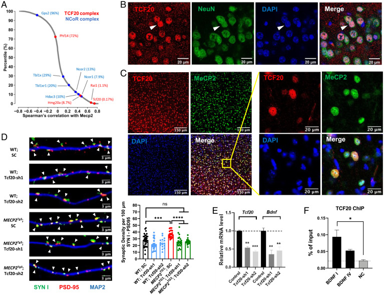
Tcf20 is coexpressed with Mecp2 in mouse neurons and regulates MECP2-mediated synapse formation. (A) Scatter plot shows the distribution of Spearman’s correlation with Mecp2 in adult mouse brain for all the genes expressed in at least one cell type. Red and blue dots denote the TCF20 complex and NCoR1/2 complex components, respectively, with top percentiles shown in parentheses. (B) Representative immunocytochemical images of mouse cortex showing TCF20 (red) is localized in the nucleus (blue, detected by DAPI) of NeuN+ (green) neurons, arrowheads. (C) Representative immunocytochemical images of mouse cortex showing TCF20 (red), MeCP2 (green), and DAPI (blue) proteins. (D) Representative images (Left) and quantification (Right bottom) of synaptic density marked by colocalization of Synapsin I (SYN I, green) and PSD-95 (red) puncta, arrowheads, with MAP2 (blue) in cultured mouse hippocampal neurons from WT and MECP2Tg3 mice infected with a nontargeting scramble (SC) AAV virus or AAV-shRNAs targeting TCF20 (n = 15 to 31, one-way ANOVA with post hoc Tukey’s tests). (E) Quantification of Tcf20 and Bdnf mRNA levels by qRT-PCR upon knockdown of Tcf20 in cultured hippocampal neurons from WT mice (n = 3, two-way ANOVA with post hoc Tukey’s tests). (F) ChIP with anti-TCF20 on cortex chromatin from WT mice. Three primer sets were used to amplify Bdnf promotor I, promoter IV, and an intergenic region as negative control (NC) (n = 3 per group, one-way ANOVA with post hoc Tukey’s tests). *P < 0.05, **P < 0.01, ***P < 0.001, ****P < 0.0001; data are mean ± SEM.

