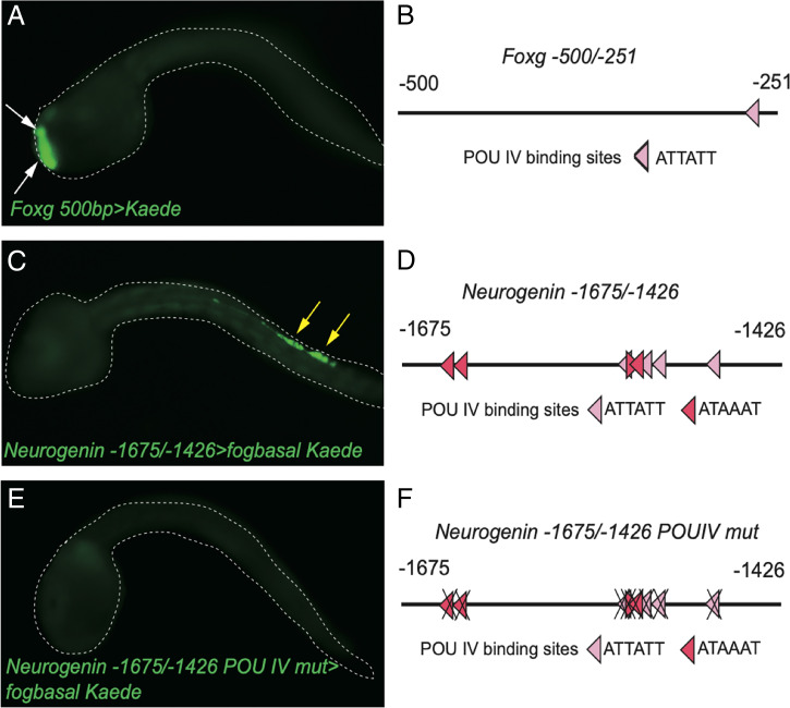
Fig. 3.
POU IV binding sites are present in the PSC minimal enhancer region of Foxg and BTN minimal enhancer region of Neurogenin and are necessary for the expression of Neurogenin in BTNs. (A) Kaede fluorescence in Foxg 500bp>Kaede–injected embryos. White arrows indicate Kaede fluorescence in PSCs. (B) Schematic diagram of -500/-251 bp of the PSC-specific Foxg minimal enhancer. One POU IV binding site is present. (C) Kaede fluorescence of Neurogenin -1675/-1472>Kaede–injected embryos. Yellow arrows indicate Kaede fluorescence in BTNs. (D) Schematic diagram of -1675/-1472 bp of the BTN-specific minimal enhancer. Eight POU IV binding sites are present. (E) Mutation analysis of eight POU IV binding sites showed that these POU IV binding sites are necessary for expression of Neurogenin in BTNs. (F) Schematic diagram of mutation analysis in E. Dark red binding sites are the higher-affinity POU IV binding sites.

