
Fig. 2.
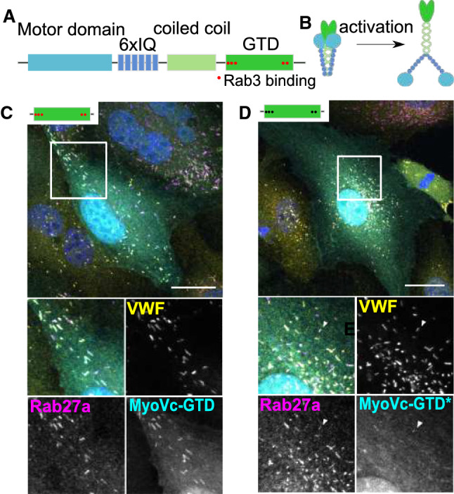
MyoVc localization to WPB is regulated by interaction with Rab3. A Depicts the domain structure of MyoVc consisting of a motor domain, 6 IQ domains, a coiled coil domain that drives dimerization, and a globular tail domain (GTD) which facilitates binding to Rab3. B Cartoon illustrating that MyoVc forms dimers that unfold upon (cargo-dependent) activation. C, D HUVEC expressing different GFP-tagged MyoVc mutants (cyan) were co-stained for VWF (yellow) and Rab27a (magenta). DAPI stainings are shown in blue. Scale bars: 10 µm. The boxed areas are shown enlarged beneath the merge. C MyoVc-GTD. D MyoVc-GTD* mutant deficient in Rab3-binding. The arrowhead depicts a WPB weakly stained for MyoVc-GTD*